Prostata MRI
Dear patient,
Dear relatives and interested parties!
Worldwide, prostate carcinoma (PCa) is the second most common cancer and the fifth most common cause of cancer death in men. In Germany, around 69,000 men are newly diagnosed with prostate cancer every year. This corresponds to around 103 out of 100,000 men or, in a city like Cologne, around 550 men per year. Around 88% of men are still alive 10 years after their initial diagnosis.
Examinations for PCa by testing the PSA value (prostate-specific antigen from the blood), ultrasound and a digital rectal examination (DRU) aim to recognise PCa at an early stage.
If prostate cancer is suspected, our task is to use MRI to visualise a tumour or find other causes for the increase in PSA. In the event of an abnormal DRU or an elevated PSA value, an mpMRI should be performed in accordance with the current urological guidelines and a decision should then be made as to whether a sample (biopsy) should be taken.
Multi-parametric magnetic resonance imaging (mpMRI) is the most precise method for detecting prostate cancer and can show the urologist the exact location of the tumour and its spread.
The combination of mpMRI and ultrasound = fusion biopsy finds over 90% of relevant prostate carcinomas and multiparametric MRI also offers the possibility of defining patient groups in future that no longer require an immediate biopsy. This means that in future it may be possible to dispense with unnecessary sampling.
The MRI examination also helps to regularly monitor patients with a less aggressive tumour (active surveillance) and to initiate treatment if the tumour progresses. In order to improve prostate diagnostics for you, we as radiologists work closely with your urologist, experienced GPs and pathologists in a network.
Pain-free examination procedure
The mpMRI examination takes approx. 35 minutes. You lie comfortably on your back during the examination. For the examination of the prostate, the patient is given a well-tolerated MRI contrast agent and a drug to calm bowel movements during the examination. Nothing needs to be inserted into the rectum.
You are welcome to bring music of your choice.
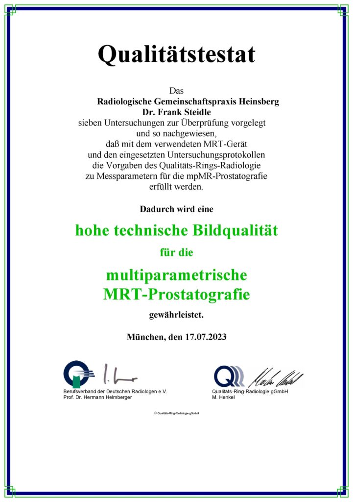
You will have the opportunity to discuss the results of the examination with the radiologist afterwards. The findings are standardised according to PI-RADS, an internationally established uniform form. This determines the probability of prostate cancer in your examination.
Costs
As we believe that this examination should be available to as many men as possible, we offer it to patients with statutory health insurance at the lowest price according to the German Medical Fee Schedule (GOÄ). Some statutory health insurance companies now cover the costs of this examination. We have a special health insurance licence for this. We will be happy to provide you with a cost estimate for the examination, which you can submit to your health insurance company. You can find the list of statutory health insurance companies that already cover the examination here: https://mediqx.de/mpMRTProstata_TeilnehmendeKrankenkassen.pdf
Private health insurance companies generally cover the costs.
Prostata MRI
ALL INFORMATION AT A GLANCE
Technology
Safety
Areas of application
Image quality
Patient comfort
We do not use an endorectal coil, i.e. no coil is inserted into the rectum. Our state-of-the-art device makes it possible to dispense with this without compromising image quality.
PROSTATE MRI PROCEDURE
Das Prinzip kurz erklärt: Die Prostata-MRT verwendet Magnetfelder und Radiowellen, um detaillierte Bilder der Prostata zu erzeugen. Der Patient liegt auf einer Liege im MRT-Gerät, und die erhaltenen Signale werden in Bilder umgewandelt.
Before examination
You will receive an information sheet before the examination. Please inform the medical staff before the examination and before entering the MRI room if you have any metallic foreign materials in your body.
In addition to the devices and implants listed below, this also applies to metal splinters, for example.
In particular, implants of the inner ear, pain and insulin pumps, pacemakers, event recorders, etc. must be accompanied by an implant ID card for written confirmation of MRI suitability prior to the examination. As metal parts in the magnetic field can cause accidents, please remove the following items before entering the examination room:
- Watch, jewelry, glasses, braces, hearing aid, metal parts on clothing (e.g. belt buckles)
- Cards with magnetic strips (e.g. check, telephone, insurance card), otherwise they will be deleted
- Keys, coins, hair clips or other objects containing metal
During the examination
- During the examination, the medical staff have visual contact with the patient through a glass pane. You are in safe hands with us.
- The duration of the examination varies and usually takes between 10 and 30 minutes; complex examinations (with contrast medium administration) can take up to 45 minutes in exceptional cases.
- During the MRI, you will lie on a couch that is moved into the tubular device.
- It is extremely important to lie still during the examination to ensure good image quality.
- The entire examination is monitored by medical staff.
- During the MRI, loud knocking noises are generated by rapidly changing magnetic fields. Patients are given headphones with music to reduce the noise.
- Depending on the problem, the medical staff may administer an intravenous contrast agent or medication during the examination.
After the examination
- The findings and further procedure will be discussed with you directly after the examination.
- The written findings will be prepared promptly and sent to your treating physicians.
