Contact
Stiftsstraße 21
52525 Heinsberg
Phone +49 (0)2452 / 924450 Fax +49 (0)2452 / 9244579
info radiologie-heinsberg.de

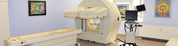
Nuclear medicine
Thyroid diagnostics
Thyroid complaints are common, and many people do not realise that their thyroid is too big, has lumps or does not work properly.
A nationwide research study revealed that 33,2% of the population has a struma (thyroid-enlargement) or a lump.
Other studies revealed that up to 20% of the population suffers from a deficient thyroid function without knowing it.
The goal of the examination is the differentiation of thyroid lumps or malfunctions of the thyroid. The doctor or assistant will therefore inject a very small amount of radioactive material into a vein on the arm. This material will nearly completely get into the thyroid. After 20 minutes, enough material will have reached the thyroid, for the examination to start. This will last 5 minutes. During this time the head must be held still to ensure that pictures taken are not blurred. Breathing and swallowing is of course allowed.
Skeleton scintigraphy
This examination is a very sensitive method to determine diseases of the bone apparatus.