DEXA bone density measurement

In our radiology department in Heinsberg, we offer the only recognised and reliable technique for determining bone density.
With advancing age, there is naturally a decrease in bone mass and therefore also in bone density. We use Dual Energy X-Ray Absorptiometry (DEXA) to recognise the risk of bone fractures at an early stage, particularly in people with reduced bone density.

The DEXA technique, also known as osteodensitometry, DXA or DPX, is a medical-technical procedure for determining bone density or the calcium salt content of bones. This procedure, which is recommended by medical associations worldwide, is considered the most reliable for assessing bone strength and fracture risk.

Bone density measurement using DEXA is a fast, precise and non-invasive diagnostic tool for detecting osteoporosis and osteopenia (reduction in bone density before osteoporosis occurs).

DEXA bone density measurement
ALL INFORMATION AT A GLANCE

Technology

Diagnostic X-ray procedure using different X-ray energy levels. Imaging of the femoral neck and lumbar spine. Computer-aided evaluation.

Safety

Low radiation dose compared to standard X-rays. Areas of application

Areas of application

Gold standard for detecting, monitoring the progress and treatment of osteoporosis and osteopenia.

Image quality

Provides quantifiable, reproducible data on bone density and thus for risk assessment for bone fractures.

Patient comfort

The examination is quick and painless. You lie comfortably on your back. The examination and analysis only take a few minutes. You will be informed of the results immediately afterwards.

Procedure

Before examination

You will receive a medical history form. Please answer the questions carefully. Based on the answers, we can better assess the fracture risk together with the measurement.
  • Medication:
Inform our medical staff about any medication and dietary supplements you are taking, as some substances can affect bone density.

During the examination
The following points must be observed during the bone density measurement using DEXA:

  • Instructions
We try to make the examination as pleasant as possible for you, but we need your co-operation to produce the best possible images. You will receive instructions and support from our staff for this purpose. Please follow these instructions as closely as possible to ensure the best possible quality of the images and to avoid repeat images.
  • Positioning:
You will lie on a comfortable examination couch. The part of the body to be examined - usually the hip and/or spine - will be positioned correctly to ensure accurate measurements.
  • Keep still:
It is important to lie still during the measurement in order to obtain clear and precise images.
  • Short duration:
The measurement only takes a few minutes.

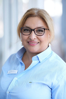
Mardjan Raptis

Specialist in nuclear medicine

Vita:

  • Born in Persia
  • Studied chemistry at the RWTH Aachen up to the intermediate diploma
  • Studied human medicine at RWTH Aachen University (UK Aachen)
  • Specialist in nuclear medicine since 2012 and in private practice
  • 2012-2017 in the group practice Radiology Aachen Land
  • 2017-2020 in the practice ZRN Grevenbroich
  • 020-03/2022 in the group practice Radiology Heinrichallee
  • Owner of the nuclear medicine practice in Heinsberg since 04/2022
  • 2 adult daughters
  • Languages: German, Persian, English

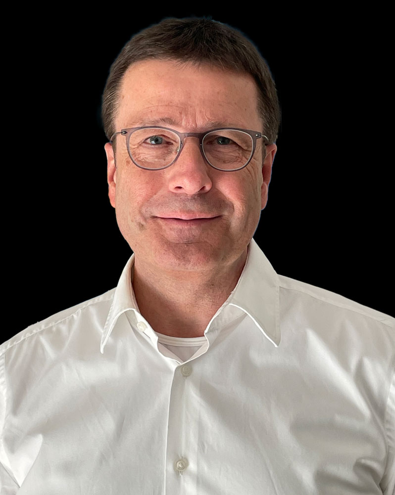
Dr. med.Werner Ihm

Specialist in diagnostic radiology

Dr Werner Ihm, born in Euskirchen, studied medicine at RWTH Aachen University from 1980-1987 and obtained his doctorate in 1988. After qualifying as a specialist in diagnostic radiology in 1997, he set up a joint practice with Dr Fernand Honinx in Heinsberg on 1 July 1999.

He specialises in magnetic resonance imaging, computer tomography and CT-guided pain therapy.
He is married and has three adult children.

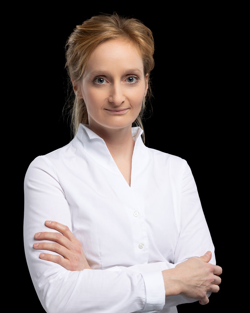
Dr. med. Inga Bosse

Specialist in diagnostic radiology

Dr. Bosse was born and grew up in Düren.

After studying human medicine at RWTH Aachen University and completing her doctorate at the Institute of Neuroradiology at RWTH Aachen University in 2013, she continued her medical training in Stolberg, Düren and Aachen.

In 2019, she practised as a radiology specialist in Aachen and since 2020 in Heinsberg.

In 2023, she took over the practice from Dr (B) Honinx in Heinsberg and in the Aachen-Düren-Heinsberg mammography screening unit.
Her main areas of expertise as a specialist are cross-sectional imaging diagnostics, CT-guided pain therapy and breast diagnostics.