Magnetic resonance imaging
(MRI)
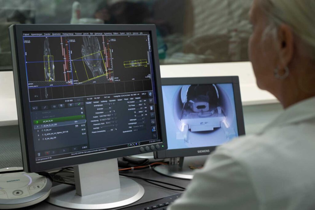
In our radiology department, we offer examinations using magnetic resonance imaging. This procedure, also known as magnetic resonance imaging (MRI for short), enables the creation of three-dimensional images without exposure to radiation. In contrast to computer tomography (CT), magnetic resonance imaging works with magnetic fields. We work with the latest standards from Siemens in order to minimise examination times while maintaining excellent image quality.
Magnetic resonance imaging (MRI)
ALL INFORMATION AT A GLANCE
Technology
Use of magnetic fields for detailed imaging of a body region.
Safety
Does not use X-rays, therefore no danger from ionizing radiation.
Areas of application
Versatile, with and without contrast medium.
Image quality
Produces high-resolution images with good differentiation of soft tissue.
Patient comfort
We try to make the examination as comfortable as possible for you. You lie on a comfortable couch and can listen to music during most examinations. A special silent mode (quiet device) further reduces the volume of the machine. State-of-the-art computers shorten the examination time while improving image quality.
Contrast medium
Occasionally it is necessary to administer a contrast medium into the vein or to drink. Occasional use to improve diagnostics, both are generally well tolerated.
We also use liver-specific contrast media if your referring physician has asked us to do so. You must register separately for this examination.
PROCEDURE OF MAGNETIC RESONANCE IMAGING (MRI)
A brief explanation of the principle: magnetic resonance imaging (MRI) uses magnetic fields and radio waves instead of X-rays, which means that patients are not exposed to radiation. The MRI machine generates a strong but basically harmless magnetic field.
As different types of tissue contain different amounts of hydrogen nuclei, they generate different signals. This makes it possible to distinguish between different types of tissue on the MRI images.
Before examination
You will receive an information sheet before the examination. Please inform the medical staff before the examination and before entering the MRI room if you have any metallic foreign materials in your body. In addition to the devices and implants listed below, this also applies to metal splinters, for example. In particular, implants of the inner ear, pain and insulin pumps, pacemakers, event recorders, etc. must be accompanied by an implant ID card for written confirmation of MRI suitability prior to the examination. As metal parts in the magnetic field can cause accidents, please remove the following items before entering the examination room:- Watch, jewelry, glasses, braces, hearing aid, metal parts on clothing (e.g. belt buckles)
- Cards with magnetic strips (e.g. check, telephone, insurance card), otherwise they will be deleted
- Keys, coins, hair clips or other objects containing metal
During the examination
- During the examination, the medical staff have visual contact with the patient through a glass pane. You are in safe hands with us.
- The duration of the examination varies and usually takes between 10 and 30 minutes; complex examinations (with contrast medium administration) can take up to 45 minutes in exceptional cases.
- During the MRI, you will lie on a couch that is moved into the tubular device.
- It is extremely important to lie still during the examination to ensure good image quality.
- The entire examination is monitored by medical staff.
- During the MRI, loud knocking noises are generated by rapidly changing magnetic fields. Patients are given headphones with music to reduce the noise.
- Depending on the problem, the medical staff may administer an intravenous contrast agent or medication during the examination.
After the examination
- If intravenous contrast medium was used, it may be helpful to drink sufficient fluids (water, tea) after the examination in order to eliminate the medium from your body via the kidneys.
- The MR images will be evaluated by a specialist radiologist after the examination. The written findings will be prepared promptly and sent to your referring doctor. Your attending physician will inform you of the results and the next steps.
