Computed tomography (CT)
Computed tomography (CT)
ALL INFORMATION AT A GLANCE
Technology
Safety
Areas of application
Image quality
Patient comfort
Precision
COMPUTED TOMOGRAPHY (CT) PROCEDURE
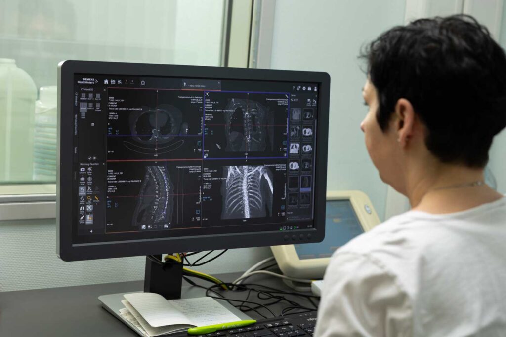
A brief explanation of the principle: CT combines state-of-the-art X-ray technology with advanced computer technology to enable fast, accurate and reliable diagnostic imaging. The rapid rotation of an X-ray tube around the patient produces detailed 3D images that enable a precise diagnosis.
Before examination
Here is some information that you should consider before the examination: You will receive an information sheet before the examination:
- If you are pregnant or suspect that you are pregnant, be sure to inform the medical staff, as computer tomography can only be performed to a limited extent and with particular caution due to the use of ionising radiation. Your doctor can recommend alternative examination methods that do not expose the unborn child to radiation.
- Allergies and medication:
Allergies and medication: Inform the medical staff of any known allergies and any prescribed medication.
- Fasting:
As a rule, it is not necessary to fast. However, this is usually necessary for abdominal examinations. This means not eating or drinking anything beforehand. You can take your medication as usual with a sip of water.
Please inform us if you suffer from diabetes or diabetes, as special rules apply here.
- Intravenous contrast medium:
Depending on the question and suspected diagnosis, it may be necessary to inject contrast medium via a venous access before or during the examination. You will be informed about this before the examination. It is important that your thyroid gland and kidneys are healthy. It is necessary that you have your TSH and creatinine levels (GFR) determined by a blood test before the examination and bring the results of the laboratory analysis with you to the examination appointment. The TSH can be dispensed with if your referring doctor confirms in writing on the referral that there is no suspicion of thyroid disease.
- Oral contrast medium:
If the abdomen is being examined, it may be necessary to drink contrast medium beforehand. For this purpose, you will be given a contrast agent diluted in water before the examination, which you should take in small sips over a period of 30 to 90 minutes (depending on the examination and the question being asked).
During the examination
The following points must be observed during the CT scan:
- Positioning:
For the examination in the computerised tomography scanner, you lie on a comfortable couch that moves slowly through the ring-shaped device. The opening is wide and the machine is open at the front and back so that you do not feel cramped. You can speak to the CT team X-ray staff at any time if you feel uncomfortable.
- Keep still and breathing instructions:
We try to make the examination as comfortable as possible for you, but we need your co-operation in order to take the best possible images. You will receive instructions and support from our staff. You will be asked to lie still and may be given instructions on breathing. Please follow these instructions as closely as possible to ensure the optimum quality of the images and to avoid repeat images. Please follow the instructions of the medical staff.
- If an intravenous contrast medium is used:
During injection, the body may experience a slight, harmless feeling of warmth as well as a strange taste in the mouth. Both will disappear after a few seconds. Patients rarely experience the feeling of warmth as discomfort or as a kind of nausea.
After the examination
- If intravenous contrast medium was used
Iit may be helpful to drink sufficient fluids (water, tea) after the examination in order to eliminate the medium from your body via the kidneys.
- Analysis of the CT images
The CT images are analysed by a radiology specialist immediately after the examination.
- Emergencies, acute or life-threatening diagnoses
In the case of emergencies, acute or life-threatening diagnoses, a medical consultation will take place immediately and further treatment will be initiated, if necessary on an inpatient basis. The written findings will be prepared immediately and sent to your referring doctor. Your attending physician will inform you of the results and the next steps.
Metal artefact reduction (iMAR reconstruction)
We offer metal artefact reduction, also known as iMAR (iterative Metal Artifact Reduction), with our state-of-the-art computer tomograph from Siemens. Metal artefact reduction is a new technology in computed tomography (CT). It aims to reduce image interference in the examination area caused by metal objects (joint implants, pacemakers, dental material, etc.) in the patient's body.
Complex image processing algorithms detect and minimise these artefacts in the iMAR reconstructions to produce clearer and more precise CT images without increased radiation exposure.
This enables more accurate diagnoses for people with implants or metallic objects in their bodies.


