Mammography

We offer digital mammography in our radiology department. This special X-ray examination of the breast is designed to detect changes at an early stage. Mammography includes two images of each breast as standard and is considered the gold standard for detecting breast cancer worldwide, even at a stage when the cancer is not yet present as a palpable lump.
During the examination, the breast is carefully but firmly positioned in a holding device and briefly compressed. This compression serves to minimise radiation exposure and maximise image quality.
Although this procedure can be uncomfortable, it is usually painless.

Mammography:
ALL INFORMATION AT A GLANCE

Technology

Digital mammography with our latest generation mammography device from Siemens for the best image quality.

Safety

Generally low radiation dose compared to conventional X-ray techniques.

Areas of application

Detection and early diagnosis of breast cancer; clarification of unclear findings and symptoms, in particular lumps or pain in a breast; regular screening examinations, in particular for women with an increased risk of breast cancer.

Image quality

High resolution for detailed visualisation of breast tissue.

Patient comfort

Short examination time, trained staff for a pain-free examination.

Aftercare

Specialist evaluation of the images; if necessary, additional sonography of the breast as well as counselling and recommendations for further steps in the event of abnormal findings.

Note

As part of the mammography screening program from the age of 50, you will receive an invitation with an appointment. If you have received an invitation for mammography screening by post, please contact the mammography screening centre (floor 2 of the medical centre)!

Mammography procedure

Before examination

  • Please bring any previous images and/or previous findings with you.
  • Please do not apply any creams or ointments to your upper body or breasts. Do not use deodorants containing aluminium.
  • For male patients: If you have a lot of hair on your upper body and chest, please remove all hair/shave your chest before the examination.
  • For premenopausal female patients: Ideally, your appointment with us should take place shortly after your period, as the breasts are less sensitive at this time.

During the examination

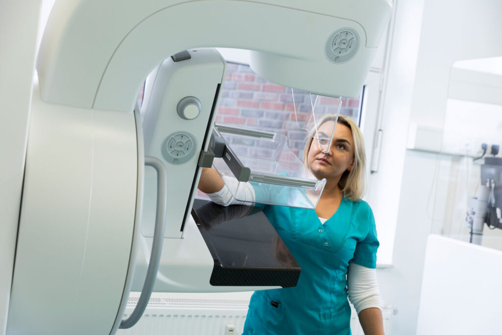
In the changing room, you will be asked to expose your upper body. In the examination room, your breast will be positioned between the mammography device in a standardised way. The breast is carefully compressed briefly to obtain clear images and minimise radiation exposure. Two images are taken per breast to ensure complete visualisation of the breast tissue.

After the examination

The examination usually takes no longer than 10 minutes. After the images have been taken, you can get dressed and take a seat in the waiting room. The images will be checked and given to you if necessary. The findings will be sent to the referring doctor promptly, usually on the same or next day.
Dear patients,

As confusion unfortunately occurs time and again, we would like to point out that we are not the mammography screening service.

So if you have received an invitation to a mammography screening, it is not from us. The screening is located in the same building, in an independent practice, two floors above us. We are neither involved in the implementation nor in the organization.

We also offer mammograms in our practice, independently of the screening.
However, we are only allowed to perform these for people with statutory health insurance on a purely curative basis, i.e. only with a valid referral from another doctor.

Mardjan Raptis

Specialist in nuclear medicine

Vita:

  • Born in Persia
  • Studied chemistry at the RWTH Aachen up to the intermediate diploma
  • Studied human medicine at RWTH Aachen University (UK Aachen)
  • Specialist in nuclear medicine since 2012 and in private practice
  • 2012-2017 in the group practice Radiology Aachen Land
  • 2017-2020 in the practice ZRN Grevenbroich
  • 020-03/2022 in the group practice Radiology Heinrichallee
  • Owner of the nuclear medicine practice in Heinsberg since 04/2022
  • 2 adult daughters
  • Languages: German, Persian, English

Dr. med.Werner Ihm

Specialist in diagnostic radiology

Dr Werner Ihm, born in Euskirchen, studied medicine at RWTH Aachen University from 1980-1987 and obtained his doctorate in 1988. After qualifying as a specialist in diagnostic radiology in 1997, he set up a joint practice with Dr Fernand Honinx in Heinsberg on 1 July 1999.

He specialises in magnetic resonance imaging, computer tomography and CT-guided pain therapy.
He is married and has three adult children.

Dr. med. Inga Bosse

Specialist in diagnostic radiology

Dr. Bosse was born and grew up in Düren.

After studying human medicine at RWTH Aachen University and completing her doctorate at the Institute of Neuroradiology at RWTH Aachen University in 2013, she continued her medical training in Stolberg, Düren and Aachen.

In 2019, she practised as a radiology specialist in Aachen and since 2020 in Heinsberg.

In 2023, she took over the practice from Dr (B) Honinx in Heinsberg and in the Aachen-Düren-Heinsberg mammography screening unit.
Her main areas of expertise as a specialist are cross-sectional imaging diagnostics, CT-guided pain therapy and breast diagnostics.